Caspase-Glo® 1 Inflammasome Assay
A Bioluminescent Method to Directly Measure Inflammasome Activity
- Fast, simple, homogeneous caspase-1 assay kit
- Selective and quantitative
- Multiplex and high-throughput screening (HTS) compatible

Catalog Number:
Size
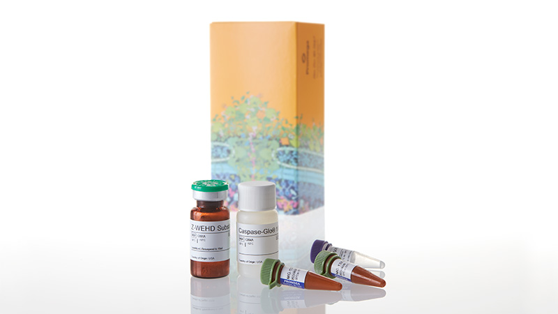
Catalog Number: G9951
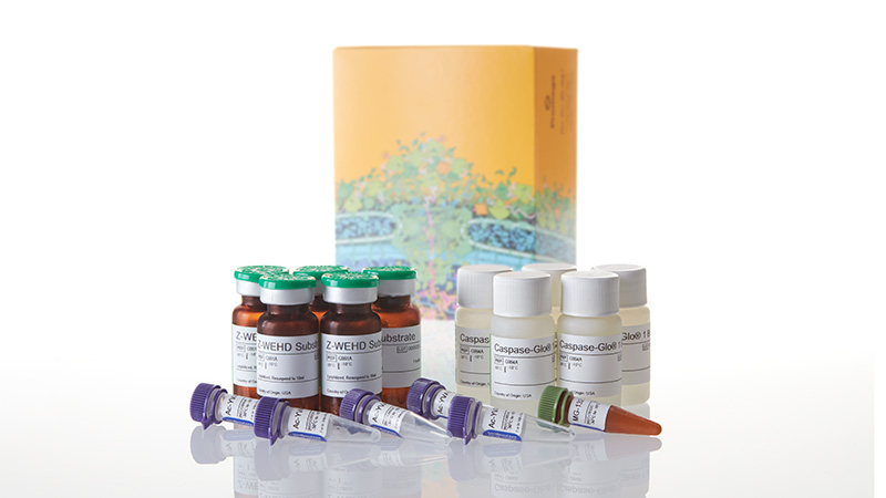
Catalog Number: G9952
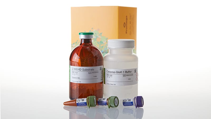
Catalog Number: G9953
Measure Caspase-1 Activity in Cells or Medium, No Sample Prep Required
The Caspase-Glo® 1 Inflammasome Assay is a simple, homogeneous, bioluminescent method to selectively measure the activity of caspase-1, an essential component of the inflammasome.
Inflammasomes are protein complexes induced by diverse inflammatory stimuli. Innate immune cells respond to pathogens and other danger signals with inflammasome formation and conversion of procaspase-1 zymogen into catalytically active caspase-1. Caspase-1 activation results in the processing and release of cytokines IL-1β and IL-18, as well as pyroptosis, an immunogenic form of cell death. The Caspase-Glo® 1 Assay is sensitive enough to measure caspase-1 activity directly in cells or in medium in multiwell plates.
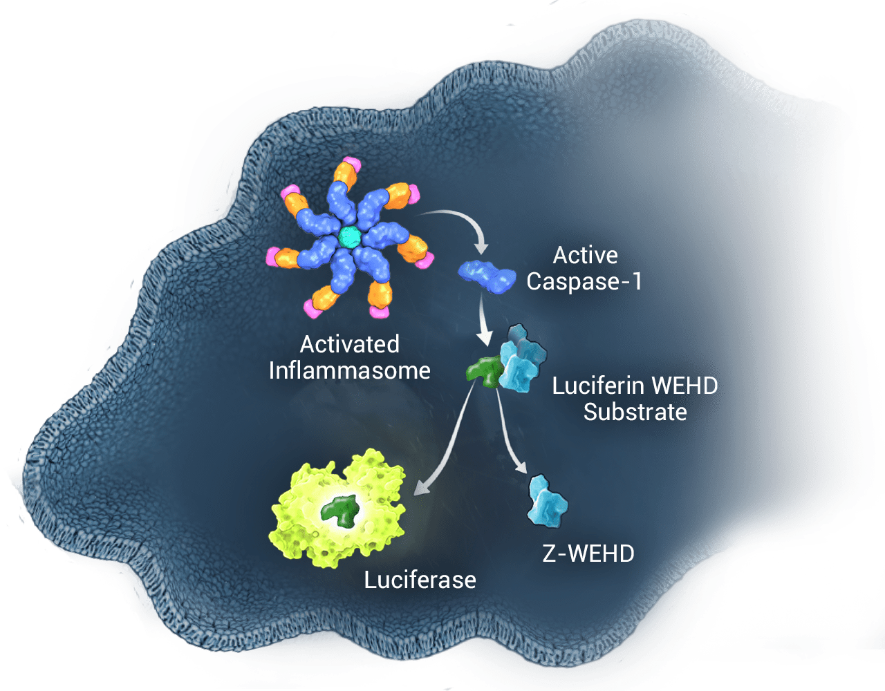
How It Works
Following caspase cleavage of the Z-WEHD substrate (Z-WEHD-aminoluciferin), a substrate for luciferase (aminoluciferin) is released, resulting in luciferase activity and generation of light by a proprietary, thermostable, recombinant luciferase.

Fast and Simple
No sample preparation or manipulation required. Simply add, mix and then measure luminescence after only one hour. You can get results faster in fewer steps compared to conventional Western blot and ELISA analyses. The stable luminescent signal and large volume (100ml) format supports high-throughput screening applications.

Selective and Quantitative
The selective caspase-1 substrate (Z-WEHD) enables detection of catalytically active caspase-1 in cells or culture media and quantitative measurement of inflammasome activity. Inclusion of the proteasome inhibitor MG-132 in the reagent eliminates non-specific proteasome substrate cleavage, enabling sensitive detection of caspase-1. The assay includes a caspase-1 specific inhibitor (Ac-YVAD-CHO) to confirm specific activity in parallel samples and distinguish caspase-1 activity from other caspases.
Caspase-1 Activity is Distinguished From Other Caspases
Inflammation and apoptosis caspases at equimolar concentrations were tested with the assay, +/– Ac-YVAD-CHO, and caspase activity was measured after 20 minutes. Ac-YVAD-CHO at 1µM inhibited 99% of the caspase-1 activity, whereas it had minimal effect on the other cross-reacting caspases (5, 3 and 6).

NLRP3 Inhibitor Prevents Caspase-1 Activation in THP-1 and PBMC Cells
NLRP3 plays a key role in immune sensing by initiating the assembly of an inflammasome in response to various danger signals. The NLRP3 inflammasome triggers caspase-1 activation and IL-1β cytokine secretion, eventually resulting in an inflammatory, pyroptotic cell death. The Caspase-Glo® 1 Assay allows you to easily measure the effect of NLRP3 inhibitors on caspase-1 activation in primary cells.

THP-1 cells were differentiated for 3 days with PMA. Medium was replaced and MCC950 (NLRP3 inflammasome inhibitor), was added followed by LPS or vehicle. Half of the culture medium (50µl) was removed after 3 hours of LPS and tested with Caspase-Glo® 1 Reagent or Caspase-Glo® 1 Reagent + YVAD-CHO.

PBMCs were plated, then treated with MCC950 followed by LPS or vehicle overnight. Following the overnight treatment, cells were treated with nigericin or vehicle. Nigericin causes potassium efflux and significant caspase-1 activation after LPS priming. Half of the culture medium (50µl) was then removed and tested with Caspase-Glo® 1 Reagent or Caspase-Glo® 1 Reagent + YVAD-CHO. Luminescence was read for both after 1 hour. Caspase-Glo® 1 signal demonstrates a dose-dependent inhibition of caspase-1 activity by the NLRP3 inflammasome inhibitor.
Caspase-1 Activity Detected in Peripheral Blood Mononuclear Cells and Released into Medium
Caspase-Glo® 1 Inflammasome Assay monitors caspase-1 activity in peripheral blood mononuclear cell (PBMC) supernatant. Cells were stimulated with or without LPS then subsequently stimulated with ATP for 30 minutes. Cells were then pelleted and supernatant was assayed for caspase-1 activity.

Multiplex With Other Cell-Based Assays
The Caspase-Glo® 1 Assay is sensitive enough to detect caspase-1 activity from a small volume of transferred medium. This leaves cells intact for other assays, such as cell viability or cytotoxicity, so you can get more data per sample.


Caspase-Glo® 1 Inflammasome Assay Multiplexed with Cell Viability and Cell Death Assays. THP-1 cells were grown in RPMI 1640 medium supplemented with 10% FBS in a 37°C incubator with 5% CO2. Cells were added to plates at 5 x 10⁵ cells/ml in 100µl of medium and differentiated with PMA (20nM, 3 days) in 96-well plates followed by treatment with flagellin (1µg/ml, 1 hour) or nigericin (20µM, 2 hours). In Panel A CellTox™ Green Reagent, Caspase-Glo® 1 Reagent or Caspase-Glo® 1 YVAD-CHO Reagent was added to the cells, and fluorescence (CellTox™ Green) or luminescence (Caspase-Glo® 1) was recorded after 90 minutes. In Panel B cell viability was monitored with CellTiter-Glo® or RealTime-Glo™ MT Cell Viability Assay. Luminescence was recorded at 10 minutes and 90 minutes, respectively.
Featured Publications
This peer-reviewed paper "A bioluminescent caspase-1 activity assay rapidly monitors inflammasome activation in cells" shows how the Caspase-Glo® 1 Assay can specifically detect caspase-1 activity in cells treated with inflammasome inducers, confirm one-signal and two-signal inflammasome activation mechanisms, and monitor caspase-1 activity, cell death, and IL-1β release from the same microplate well.
This peer-reviewed study "Interrogating direct NLRP3 engagement and functional inflammasome inhibition using cellular assays" examines the direct engagement and functionality of NLRP3 pathway activity in cells using several mechanistic assays, including the Lumit® IL-1β Immunoassay and Caspase-Glo® 1 Inflammasome Assay.
Protocols
Complete Protocol
Specifications
Catalog Number:
Contenido
| Item | Part # | Presentación | Concentración |
|---|---|---|---|
|
MG-132 Inhibitor |
G932B | 2 × 30μl | |
Z-WEHD Substrate |
G991A | 1 × 1 bottle | |
Ac-YVAD-CHO |
G992A | 1 × 10μl | 2mM |
Caspase-Glo® 1 Buffer |
G994A | 1 × 10ml |
SDS
Search for SDSCertificado de Análisis
Use Restrictions
For Research Use Only. Not for Use in Diagnostic Procedures.Condiciones de Almacenaje
Contenido
| Item | Part # | Presentación | Concentración |
|---|---|---|---|
|
MG-132 Inhibitor |
G932C | 1 × 300μl | |
Z-WEHD Substrate |
G991A | 5 × 1 bottle | |
Ac-YVAD-CHO |
G992A | 5 × 10μl | 2mM |
Caspase-Glo® 1 Buffer |
G994A | 5 × 10ml |
SDS
Search for SDSCertificado de Análisis
Use Restrictions
For Research Use Only. Not for Use in Diagnostic Procedures.Condiciones de Almacenaje
Contenido
| Item | Part # | Presentación | Concentración |
|---|---|---|---|
Caspase-Glo® 1 Buffer |
G994B | 1 × 100ml | |
|
MG-132 Inhibitor |
G932C | 2 × 300μl | |
Z-WEHD Substrate |
G991B | 1 × 1 bottle | |
Ac-YVAD-CHO |
G992B | 1 × 100μl | 2mM |
SDS
Search for SDSCertificado de Análisis
Use Restrictions
For Research Use Only. Not for Use in Diagnostic Procedures.Condiciones de Almacenaje
Resources
Artículos
- Nota de Aplicación: Cultivo de células THP-1 para ensayos de activación del inflamasoma
- Overcoming preclinical safety obstacles to discover GDC-2394: A potent and selective NLRP3 inhibitor
- High-pressure oxygen rewires glucose metabolism of patient-derived glioblastoma cells and fuels inflammasome response
Related Products
Productos Similares
Lumit® IL-1β Human/Mouse Immunoassay
Cuantifica la activación del inflamasoma mediante la medición de la IL-1β liberada utilizando un protocolo sencillo y sin lavado.
W6010, W6011, W6012, W7010, W7011, W7012, W116A-C, W119A-C
Lumit® Active IL-18 (Human) Immunoassay
Mide cuantitativamente la IL-18 activa en muestras de cultivos celulares mediante un protocolo sencillo y sin lavado que se completa en 70 minutos o menos.
W1910, W1911, W1912, W191A-C
CellTox™ Green Cytotoxicity Assay
Mide los cambios en la integridad de la membrana. Monitoriza cinéticamente la citotoxicidad hasta 72 horas y además tiene capacidad multiplex.
G8741, G8742, G8743, G8731
CellTiter-Glo® 2.0 Cell Viability Assay
Versión actualizada del CellTiter-Glo® Cell Viability Assay con una estabilidad del reactivo mejorada. Cuantifica la proliferación celular basándose en la detección de ATP.
G9241, G9242, G9243
Usado con frecuencia junto con
GloMax® Discover System
Lector de microplacas de alto rendimiento para la detección de luminiscencia, fluorescencia y absorbancia.
GM3000
Caspase-Glo® 3/7 Assay System
Ensayo luminiscente fácil de usar que permite detectar la actividad de la caspasa-3/7.
G8090, G8091, G8093, G8092
LDH-Glo™ Cytotoxicity Assay
Ensayo LDH basado en bioluminiscencia para detectar de forma sensible la citotoxicidad en muestras con bajo número de células, incluidos los microtejidos 3D.
J2380, J2381
Lumit® TNF-α (Human) Immunoassay
Mide cuantitativamente el TNF-α liberado en muestras de cultivos celulares mediante un protocolo sencillo y sin lavado.
W6050, W6052, W6051, W137A-C